When it comes to research, scientific breakthroughs are often found in the atomic details.
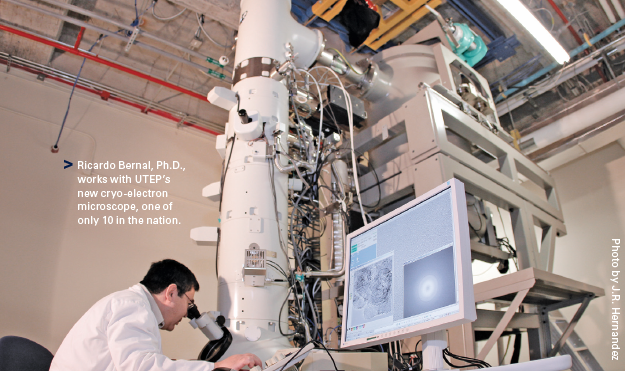
Ricardo Bernal, Ph.D., is used to looking at the tiniest of samples – subcellular images of biological materials – while exploring research applications that have implications for future cancer treatments.
The assistant professor of chemistry at The University of Texas at El Paso is paying particular attention to chaperonins. These protein complexes are found in the cytoplasm of cells and are responsible for refolding other proteins to make them functional again. But what would happen if the chaperonin failed to function?
“Cancer cells are not the healthiest cells,” Bernal said. “So as they continue to divide rapidly, they’re a mess. The chaperonin’s function inadvertently helps cancer cells survive by refolding their hastily synthesized proteins. Without a functional chaperonin, a cancer cell would die.”
This study of life at the basic level uses four complementary techniques: X-ray protein crystallography, nuclear magnetic resonance spectroscopy (NMR), cryo-electron microscopy (cryo-EM) and bioinformatics – all now available at UTEP.
The newest instrument is the $1.8 million cryo-electron microscope – funded in large part by a National Science Foundation Grant – housed in the new Chemistry and Computer Science Building.
“We’re in a unique position, having this high-end microscope, since there are only about 10 like it in the nation and only about 40 in the world,” said Bernal, chief investigator for the National Science Foundation Major Research Instrumentation grant that helped purchase the microscope. “We anticipate having users from all over the country collaborating with us and visiting the facility.”
Using the massive piece of equipment to view things at nearly an atomic level has huge implications for research.
In the past, Bernal’s research has focused on chaperonins as found in bacteria and viruses. With the installation of the cryo-EM at UTEP, his research will shift to human chaperonins that have been implicated in the survival of cancer cells.
The state-of-the-art instrument comes as the University is establishing a structural biochemistry research core. This is part of a larger movement by UTEP to achieve Tier One status by expanding research capabilities on campus and becoming the first national research university with a 21st century student demographic.
“The University is investing not only in the instrument but in expert faculty such as Dr. Ricardo Bernal who will be able to utilize this very sophisticated microscope to further the research achievements associated with UTEP,” said Stephen Aley, Ph.D., associate dean of the College of Science at UTEP. “These major investments are important on our road to Tier One.”
Bernal, a Texas A&M University graduate, earned his master’s degree in biological science in 1993 from UTEP. He spent three years at the National Cancer Institute, part of the National Institutes of Health in Maryland. He received his Ph.D. in biochemistry and molecular biology with an emphasis in structural biology in 2002 from Purdue University – where he worked under Michael Rossmann, a Hanley Distinguished Professor of Biological Science.
Bernal arrived as a faculty member at UTEP in 2006 by way of postdoctoral work at the Medical Research Council – Laboratory of Molecular Biology in Cambridge, England – the birthplace of structural biology and an institute that has produced more than 13 Nobel Laureates.
“The allure of coming here was the idea of setting up a cryo-EM facility,” Bernal said.
“UTEP was the most attractive offer in the sense that this is where I’d have the biggest impact. I think it was a great decision and I don’t regret it at all.”
Using the cryo-electron microscope, Bernal can create three-dimensional images of the chaperonin to aid in his research. These 3-D reconstructions can then yield critical information about the function of a particular biological molecule.
The 300-kilovolt microscope, purchased from JEOL, a Japanese corporation, uses a concentrated electron beam to penetrate unstained biological samples to produce low-contrast, two-dimensional images.
The bulky and expensive microscope is unique in that it can produce an extremely high vacuum. This vacuum allows for emission of electrons from what is called a field emission gun (FEG). The electron beam produced by a FEG is superior in that it is very intense and coherent, which in turn produces images of a higher quality.
Prior to the installation of the new microscope, Bernal would travel the country for the opportunity to use a cryo-electron microscope for his research.
“Before ours was installed, other universities were very generous in letting us use their cryo-electron microscope,” Bernal said. “I think that’s actually the way science is supposed to work.”
Sudheer Molugu, a postdoctoral researcher in chemistry at UTEP, has worked with Bernal for the last five years. Having a hand in research, including exploring the applications of structural biochemistry, Molugu is impressed with the work Bernal has accomplished. But more impressive is Bernal as a mentor.
“I would not have done a Ph.D. in chemistry if Dr. Bernal was not here,” Molugu said.